
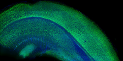
1 mm thick mouse brain labeled with NeuN and DAPI
We made our Tissue Clearing Ebook so that any researcher can easily adopt tissue clearing into their research workflow.
WHITEHOUSE STATION, N.J. (PRWEB)
August 19, 2019
Since the microscope was first introduced, researchers and clinicians have characterized tissues through the use of ultra-thin two dimensional slices which are placed on microscope slides, stained and visualized by a pathologist. However, this paradigm can be highly limited for complex tissues and complicated features such as vasculature and thus researchers in the last few years have begun to employ tissue clearing approaches combined with fluorescent labeling and three-dimensional microscopy (e.g. confocal microscopy, light sheet microscopy) to image tissues in 3D. Of all the tissue interrogation tools used in a laboratory, tissue clearing and 3D tissue imaging can be one of the most challenging and complex as it involves the marriage of multiple disparate disciplines. Proper execution of tissue clearing approaches requires an in-depth knowledge of fluorescent labeling, tissue processing, advanced microscopy, image processing and data processing.
While adopting tissue clearing can be challenging, at Visikol we have focused for the last few years on developing protocols, tools and reagents that are easy-to-use, rapid and inexpensive such that any researcher can quickly adopt them into their workflow. “To help support these efforts, we have developed a Tissue Clearing Ebook which provides a contextual overview to adopting any tissue clearing technique with feedback and lessons learned from hundreds of researchers,” described Visikol Product Manager Ian MacCloud. The Ebook is designed as a primer for adopting tissue clearing and gives researchers a break down of common pitfalls and key considerations to keep in mind during 3D imaging such as maximizing fluorescent signal, minimizing background fluorescence, when to use confocal or light sheet microscopy, which antibodies to use and how to process specific types of tissue (e.g. FFPE, frozen fresh) or 3D cell culture models (i.e. organoids, spheroids, microtisses).
“In working with lots of researchers, we have seen many of them get discouraged by tissue clearing in general as they see a 3D image of a whole mouse brain acquired with CLARITY, try to replicate the process in their lab and unfortunately have poor results. We really made the Ebook for these researchers as there are lots of mistakes researchers typically make in adopting tissue clearing such as jumping to a whole mouse brain before validating that their immunolabels can penetrate thick tissue or if they have enough fluorescent signal with thick samples,” described Visikol CSO Dr. Tom Villani. The goal of the tissue clearing Ebook is to provide a dense but short preview to tissue clearing that every researcher should review before adopting tissue clearing into their workflow. To download the Ebook click below:
TISSUE CLEARING EBOOK DOWNLOAD
About Visikol
Visikol is a CRO focused on advanced drug discovery that is leading the fields of bio-imaging, bioinformatics and image analysis. We conduct end-to-end drug discovery services that include both 2D and 3D in vitro models and assays, 3D whole mount tissue imaging, digital pathology and custom drug discovery projects. Visikol offers a portfolio of drug discovery services ranging from 2D and 3D cell culture model and assay development to in vitro screening, animal tissue histology and automated image processing. The focus of these services is to transform tissues into images and ultimately into quantitative data sets that can be mined for actionable insights that help our Clients make more informed decisions during the drug discovery process. Additionally, Visikol manufactures and sells a suite of tissue clearing reagents and 3D immuno-labeling kits. These products allow researchers to easily and rapidly image whole tissues and 3D cell culture models in 3D instead of traditional 2D sectioning. For more information about Visikol or its services, please visit our website at visikol.com.
Share article on social media or email: