Category Archives: Science: Biology

Cutting edge research is happening on a daily basis in our modern world, Keep up to date with our Press Release services.

UMD Researcher Discovers a New Mechanism in the Liver that Helps Prevent Invasive Fungal Infections


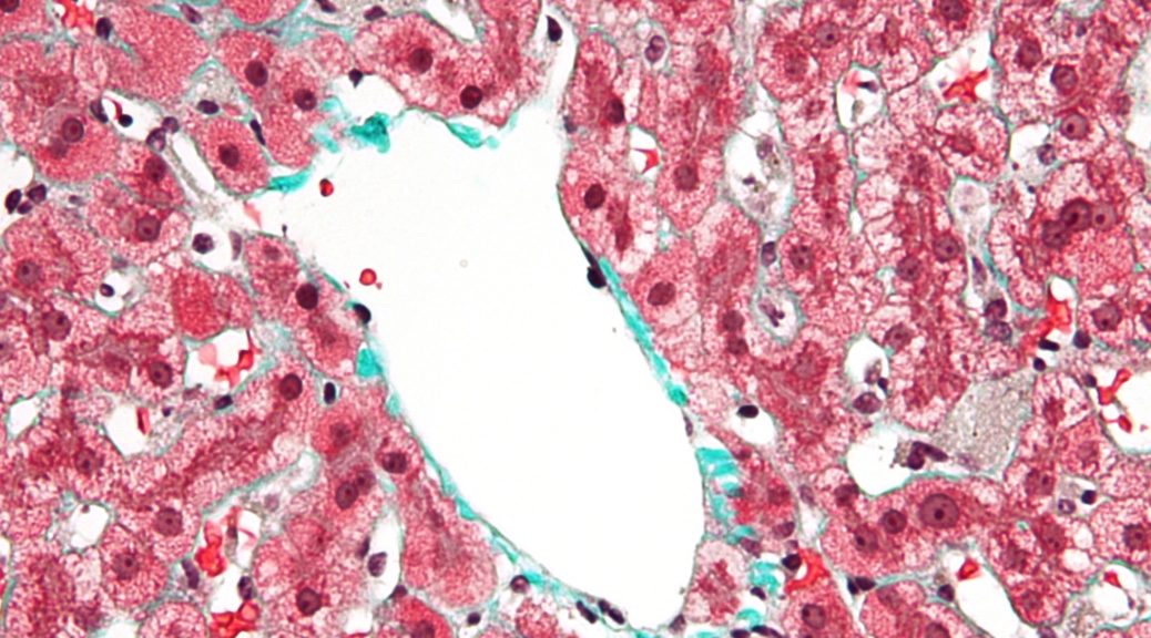
Liver macrophage cells

Liver macrophage cells, Credit: Nephron

“This is a protective mechanism that is working once the fungus becomes invasive, or gets into the bloodstream, to prevent it from spreading. Stopping the dissemination process throughout the body is so important, because once you get dissemination, you get the disease.”

As a world-renowned expert using intravital microscopy to observe fungal infection progression in live samples, Meiqing Shi, associate professor with the University of Maryland Department of Veterinary Medicine, continues to make new breakthroughs in the field. In his latest paper published in Nature Communications, Shi and his lab have discovered a new pathway by which liver macrophages called Kupffer cells (KCs) capture potentially fatal fungi in the bloodstream before dissemination to target organs like the brain or kidney. This mechanism not only provides an explanation as to why individuals with liver disease tend to have enhanced risk of contracting fungal infections, but it also points to therapeutic options that could be used in the future to prevent fungal dissemination and invasive fungal infections, which kill 1.5 million people worldwide each year.

“Under intravital microscopy, we can directly see how the KCs catch fungi in real time,” says Shi. “This is a protective mechanism that is working once the fungus becomes invasive, or gets into the bloodstream, to prevent it from spreading. Stopping the dissemination process throughout the body is so important, because once you get dissemination, you get the disease. These findings suggest therapeutic strategies for preventing dissemination, and this could be applied across many types of fungal infections, since they work in similar ways.”

Fungal infections affect 1.2 billion people globally each year. In the current paper, Shi specifically examined two types of fungi – Cryptococcus neoformans and Candida albicans. Both of these fungi, if disseminated to their target organs (the brain for Cryptococcus and the kidney for Candida), are fatal infections that are very difficult to treat once contracted. Cryptococcus, for example, is the main cause of meningitis. Each year, more than a million people are infected and contract meningitis, and 60% of those who are infected will die from the disease.

“Cryptococcus and Candida are fungi that are actually everywhere,” says Shi. “People with healthy immune systems can usually control the fungi after infection, but once it gets into the bloodstream, either one of these fungi can get into the target organs and become fatal. For Cryptococcus, this is especially a problem for those with impaired immune systems, like HIV patients or organ transplant patients. Patients with liver disease are also more prone to Cryptococcus infection, and no one understood why before.”

This new discovery that KCs or liver macrophages are responsible for catching free fungi in the bloodstream to prevent further dissemination helps explain this phenomenon, since if the liver is impaired as it is in patients with liver disease, it would stand to reason that this protective mechanism would also be impaired.

“This finding is very interesting and very unusual, because in the field of fungal infections, nobody focuses on the liver,” says Shi. “Researchers tend to look at the target organs like the brain or kidney. The liver is not a target organ, but it tries to clean out the fungus in the bloodstream. As the whole body is connected, this paper gives a more whole system approach to how fungal dissemination interacts in the entire body.”

With this whole body approach in mind, the discovery of this mechanism has implications not just for those with liver disease, but for the treatment of fungal infections as a whole by targeting this mechanism, preventing fungal dissemination, and treating invasive fungal infections.

The paper, entitled “Fungal dissemination is limited by liver macrophage filtration of the blood,” is published in Nature Communications, DOI: 10.1038/s41467-019-12381-5.

This work is funded by the National Institutes of Allergy and Infectious Diseases, National Institutes of Health, grant AI131219.

Share article on social media or email:

Research Demonstrates Impact of 2’-Fucosyllactose (2’FL) on Gut Microbiota and Metabolite Production


News Image

These kind of study approaches can give more understanding on the how HMOs can be utilized by the microbiota with a method that is not invasive, which is important especially in the case of infants.

Continuing its leading research on human milk oligosaccharides (HMOs), DuPont released the findings of a new study showing that Care4U™ 2’-FL, a DuPont product, beneficially changes microbiota composition and the production of microbial metabolites in comparison to galacto-oligosaccharides (GOS) and lactose. The effects elicited to microbiota and metabolite composition by 2’-FL were between the responses elicited by GOS and lactose, which indicates that 2’-FL is selectively fermented by the infant microbes.

“Our previous in vitro cultivation experiments, as well as literature, indicated that 2’-FL is selectively utilized by only certain types of bifidobacteria and Bacteroides species, and this type of modelling with a complex microbiota is a step ahead in our journey to understand the prebiotic mechanisms of 2’-FL,” indicated Krista Salli, M.Sc., Scientist, DuPont Nutrition & Biosciences.

“This study exemplifies the utility of the DuPont™ Enteromix® infant gut simulator to compare the fermentation of the carbohydrates between each other,” said Heli Anglenius, Ph.D., Senior Scientist, DuPont Nutrition & Biosciences. “Over the years, the Enteromix® gut simulator developed in-house in Kantvik, Finland, has been used successfully for a multitude of studies with fecal microbiota sampled from adults, and this study demonstrates its applicability to studies where the microbiota is obtained from infants. This is a great continuation for more than 20 years of expertise using our own pre-clinical modelling.”

Among the study’s other achievements was the comparison of 2’-FL to GOS and lactose.

“While this study was done under simulated gut conditions, it showed the differences between the carbohydrates, especially in the production of microbial metabolites,” said Ratna Mukherjea, Ph.D., Technical Fellow and Global Research and Development Leader-Human Milk Oligosaccharides (HMO), DuPont Nutrition & Biosciences. “These kind of study approaches can give more understanding on the how HMOs can be utilized by the microbiota with a method that is not invasive, which is important especially in the case of infants.”

You can read more about this study at https://www.nature.com/articles/s41598-019-49497-z.

About DuPont™ Danisco®

DuPont™ Danisco® is the brand for a range of products that help provide enhanced bioprotection, an improved nutritional profile, and better taste and texture with greater cost efficiency and lower environmental impact, meeting the needs of manufacturers of food and beverages and dietary supplements. Through the work of the global network of food scientists and technologists in DuPont, the Danisco® range is supported by a uniquely broad spectrum of know-how across applications and processing.

About DuPont Nutrition & Biosciences

DuPont Nutrition & Biosciences applies expert science to advance market-driven, healthy and sustainable solutions for the food, beverage, dietary supplement and pharmaceutical industries. We also use cutting-edge biotechnology across a range of markets to advance bio-based solutions to meet the needs of a growing population, while protecting our environment for future generations. We are innovative solvers who help our customers turn challenges into high-value business opportunities. For more information: http://www.dupontnutritionandbiosciences.com or http://www.biosciences.dupont.com.

About DuPont

DuPont (NYSE: DD) is a global innovation leader with technology-based materials, ingredients and solutions that help transform industries and everyday life. Our employees apply diverse science and expertise to help customers advance their best ideas and deliver essential innovations in key markets including electronics, transportation, construction, water, health and wellness, food, and worker safety. More information can be found at http://www.dupont.com.

# # #

10/08/19

DuPont™, the DuPont Oval Logo, and all trademarks and service marks denoted with ™, SM or ® are owned by affiliates of DuPont de Nemours, Inc. unless otherwise noted.

Share article on social media or email:
Asymmetrex Obtains UK Patent for Tissue Stem Cell-Specific Counting and Testing


https://www.prweb.com/

Asymmetrex Advances Stem Cell Counting Tech To UK

It was one thing to be selling products with no stem cell number or dose, when it was not possible to determine the stem cell-specific count. But it’s a whole other thing, when it is now possible. Change is here.

Today, Massachusetts stem cell biotechnology company Asymmetrex announces the recent patenting in the U.K. of its lead development technology. Like the company’s precursor patent, approved in the U.S. in 2017, the new patent covers the company’s unique technology for specific and accurate counting of tissue stem cells. Asymmetrex now markets the technology in the U.S. as the AlphaSTEM Test™. The new patent will support marketing and sales and investor development initiatives in the U.K.

Asymmetrex’s AlphaSTEM Test™ is the first and only technology to provide specific and accurate counting of tissue stem cells, both from mature adult tissues and perinatal tissues like the umbilical cord. By counting tissue stem cells specifically, the technology can determine the stem cell-specific dose of tissue stem cell treatments. The lack of this crucial information has limited progress in stem cell clinical trials that largely cannot be interpreted without it. Even currently approved stem cell treatments would benefit from stem cell-specific dose information. For example, in the case of bone marrow stem cell transplants, for which donors are scarce, knowing the stem cell-specific dose might allow additional patients to be treated with the same bone marrow donation.

Some areas of stem cell research and stem cell medicine have been slow to embrace the new technology. This is particularly true of companies that presently market isolated, manufactured, or cultured tissue stem cell-containing products, whether for research purposes or for patient treatments. Many have been unwilling to use the new technology to begin certifying the stem cell-specific count or dose of their products. Asymmetrex founder and director James L. Sherley, M.D., Ph.D., chides that this condition cannot persist. “It was one thing to be selling products with no stem cell number or dose, when it was not possible to determine the stem cell-specific count. But it’s a whole other thing, when it is now possible. Change is here.”

Specific stem cell counting is a less clouded issue for academic stem cell research labs. Basic research investigators would like to know how many stem cells are in their experiments. Accordingly, Asymmetrex is presently beta-testing the use of the technology in both U.S. and U.K. labs to position the technology to be more practical and affordable for stem cell investigators in colleges and universities.

There are two other broad categories of applications for the stem cell counting technology that often are not self-apparent. Gene therapy, including emerging gene editing, would benefit from tissue stem cell-specific counting. Because of their long-lived tissue renewing property, stem cells are the crucial targets for gene modifications to achieve single-dose, durable cures. Current clinical studies attempt gene therapy while blind to the number of stem cells during genetic modification procedures, as well as blind to the dose of genetically-modified stem cells in the final treatments.

Both Asymmetrex’s U.S. and U.K. patents cover the use of the stem cell counting technology to identify drug candidates that alter stem cells, whether with beneficial effects or detrimental effects. Drugs with beneficial effects may have efficacy for accelerating or improving tissue repair of wounds and injuries. Drug candidates with detrimental effects have even greater significance in drug development. Tissue stem cell-toxic agents cause chronic organ failures, like bone marrow or liver. This problem is estimated to be responsible for half of drug failures due to unacceptable toxicity, which are a major cause of unsuccessful drug programs. Previously, without a way to do inexpensive stem cell toxicity testing, U.S. pharmaceutical companies spent $4-5 billion each year watching these drugs fail in animal studies or subsequent clinical trials. By showing that its AlphaSTEM Test™ can identify drugs known to cause chronic organ failure, Asymmetrex is positioning itself to relieve the pharmaceutical industry of this huge cost burden with a simple, inexpensive cell culture test.

Later this month, Asymmetrex will begin a podcast series to expand and accelerate education of the inevitability of the change of stem cell research, stem cell medicine, stem cell manufacturing, and drug development to quantitative analyses for tissue stem cells. Earlier this year, the FDA Standards Coordinating Body (SCB) listed stem cell specific-dose determination as a priority for needed standards for regenerative medicine and stem cell therapies. Dose is a fundamental principle for the discovery, development, and administration of quality medicines. Sherley assures, “It can be no different for stem cell medicine. Our podcast will not just target the stem cell medicine and pharmaceutical industry. We also wish to inform the end users of stem cell products, patients, their families, and their advocates, including the FDA. When they understand and demand certification the quality and dose of stem cells in their treatments, the industry will speed up its adoption of the change that must come.”

About Asymmetrex

Asymmetrex, LLC is a Massachusetts life sciences company with a focus on developing technologies to advance stem cell medicine. Asymmetrex’s founder and director, James L. Sherley, M.D., Ph.D. is an internationally recognized expert on the unique properties of adult tissue stem cells. The company’s patent portfolio contains biotechnologies that solve the two main technical problems – production and quantification – that have stood in the way of successful commercialization of human adult tissue stem cells for regenerative medicine and drug development. In addition, the portfolio includes novel technologies for isolating cancer stem cells and producing induced pluripotent stem cells for disease research purposes. Asymmetrex markets the first technology for determination of the dose and quality of tissue stem cell preparations (the “AlphaSTEM Test™”) for use in stem cell transplantation therapies and pre-clinical drug evaluations.

Share article on social media or email:

New Study Discovers the Three-Dimensional Structure of the Genome Replication Machine


Mount Sinai researchers have discovered how the enzyme DNA polymerase delta works to duplicate the genome that cells hand down from one generation to the next. In a study published in Nature Structural & Molecular Biology, the team also reported how certain mutations can modulate the activity of this enzyme, leading to cancers and other diseases.

“DNA polymerase delta serves as the duplicating machine for the millions to billions of base pairs in human and other genomes. We were able to present for the first time a near-atomic-resolution structure of the complete enzyme in the act of DNA synthesis,” says lead investigator Aneel Aggarwal, PhD, Professor of Pharmacological Sciences at the Icahn School of Medicine at Mount Sinai. “This knowledge furthers our basic understanding of this complex enzyme which is essential for survival in higher organisms from humans to yeast. At the same time, our work provides insights into how cancers can arise when DNA polymerase delta is not functioning properly, and offers a novel basis for designing inhibitors of the polymerase that could potentially serve as effective treatment in certain cancers.”

While DNA polymerase delta has been studied by scientists for decades, many questions remain about its overall architecture and dynamics. “We showed how the various pieces of this complicated machine work synchronously with one another to copy the genome with amazing accuracy,” explains Dr. Aggarwal. His team, which included co-author Rinku Jain, PhD, Assistant Professor of Pharmacological Sciences at the Icahn School of Medicine, also mapped a number of inherited mutations (which are passed down from parent to child) and somatic mutations (which occur by chance during someone’s lifetime) in DNA polymerase delta that are associated with “hypermutated” tumors. In addition to cancers, these mutations may be associated with multi-symptom mandibular hypoplasia, deafness, and lipodystrophy syndrome.

Essential to the Mount Sinai researchers’ work were recent advances in cryo-electron microscopy. This technology, which allows for the imaging of rapidly frozen molecules in solution, is revolutionizing the field of structural biology through its high-resolution pictures. This technique allowed Dr. Aggarwal and his team to examine not only individual atoms of the DNA polymerase delta but also how they move to achieve accurate replication of the genome. Integral to this phase of the research was Mount Sinai’s partnership with the Simons Electron Microscopy Center in New York City.

Building on its latest groundbreaking work around DNA polymerase delta, Mount Sinai will continue to explore the unique structure and mechanism of the polymerase, particularly its relationship to cancer and disease pathogenesis. “We know that certain cancers become dependent on this enzyme for their survival,” says Dr. Aggarwal, “and inhibiting its activity could provide a valuable therapeutic window for future medical research.”

About the Mount Sinai Health System

The Mount Sinai Health System is New York City’s largest integrated delivery system, encompassing eight hospitals, a leading medical school, and a vast network of ambulatory practices throughout the greater New York region. Mount Sinai’s vision is to produce the safest care, the highest quality, the highest satisfaction, the best access and the best value of any health system in the nation. The Health System includes approximately 7,480 primary and specialty care physicians; 11 joint-venture ambulatory surgery centers; more than 410 ambulatory practices throughout the five boroughs of New York City, Westchester, Long Island, and Florida; and 31 affiliated community health centers. The Icahn School of Medicine is one of three medical schools that have earned distinction by multiple indicators: ranked in the top 20 by U.S. News & World Report’s “Best Medical Schools”, aligned with a U.S. News & World Report’s “Honor Roll” Hospital, No. 12 in the nation for National Institutes of Health funding, and among the top 10 most innovative research institutions as ranked by the journal Nature in its Nature Innovation Index. This reflects a special level of excellence in education, clinical practice, and research. The Mount Sinai Hospital is ranked No. 14 on U.S. News & World Report’s “Honor Roll” of top U.S. hospitals; it is one of the nation’s top 20 hospitals in Cardiology/Heart Surgery, Diabetes/Endocrinology, Gastroenterology/GI Surgery, Geriatrics, Gynecology, Nephrology, Neurology/Neurosurgery, and Orthopedics in the 2019-2020 “Best Hospitals” issue. Mount Sinai’s Kravis Children’s Hospital also is ranked nationally in five out of ten pediatric specialties by U.S. News & World Report. The New York Eye and Ear Infirmary of Mount Sinai is ranked 12th nationally for Ophthalmology and Mount Sinai South Nassau is ranked 35th nationally for Urology. Mount Sinai Beth Israel, Mount Sinai St. Luke’s, Mount Sinai West, and Mount Sinai South Nassau are ranked regionally.

For more information, visit https://www.mountsinai.org or find Mount Sinai on Facebook, Twitter and YouTube.

Share article on social media or email:

Asymmetrex Leads OCTNE 2019 Discussion of the Growing Need for Tissue Stem Cell-Specific Dosing In Stem Cell Clinical Trials


https://www.prweb.com/

Unlike all other medicines, the dose of treatment stem cells continues to be ignored.

If the FDA is going to regulate stem cells like other medicines, now that a counting technology is available, they should also guide this industry to its inevitable adoption of dose, a universally acknowledged, fundamental requirement for progress and success in medicine.

Yesterday, at the 2019 meeting of Outsourcing in Clinical Trials New England in Boston, October 2-3, Asymmetrex founder and director, James L. Sherley, M.D., Ph.D., discussed important design factors that currently limit the success of early stage stem cell clinical trials. To an attendee audience including both developers and suppliers of clinical trials, in his talk Dr. Sherley focused on the highly problematic practice of conducting stem cell clinical trials and stem cell treatments without knowing stem cell dose. He referred to the widespread lack of stem cell-specific dosing as “the major unacknowledged design feature that currently limits the success of ALL stem cell clinical trials.”

As for all effective medicines, stem cells are no different when it comes to the importance of knowing the dose. Sherley pointed out that, without knowing the dose of administered stem cells, it is impossible to interpret the outcome of current clinical trials. With an audience including manufacturers and suppliers of stem cell treatments for clinical trials, Sherley emphasized that the stem cell-specific counting problem extended to many aspects of producing and certifying the quality of tissue stem cell treatment preparations. In the case of manufacturing processes that aim to expand the number of tissue stem cells, some products may have diluted the stem cells to a negligible number and an ineffective dose.

Sherley also shared with attendees that change was now on the horizon of the regulatory landscape for stem cell-specific dosing. With the motivation of Asymmetrex’s new AlphaSTEM Test™ technology, which now makes specific and accurate counting of therapeutic tissue stem cells possible, earlier this year the FDA’s Standards Coordinating Body for Regenerative Medicine (SCB) listed stem cell-specific dose determination as a priority for the development needed standards to advance cellular therapies. Sherley notes, “This was a critical milestone for catching up stem cell medicine to being quantitative, like all the rest of medicine. If the FDA is going to regulate stem cells like other medicines, now that a counting technology is available, they should also guide this industry to its inevitable adoption of dose, a universally acknowledged, fundamental requirement for progress and success in medicine.”

About Asymmetrex

Asymmetrex, LLC is a Massachusetts life sciences company with a focus on developing technologies to advance stem cell medicine. Asymmetrex’s founder and director, James L. Sherley, M.D., Ph.D. is an internationally recognized expert on the unique properties of adult tissue stem cells. The company’s patent portfolio contains biotechnologies that solve the two main technical problems – production and quantification – that have stood in the way of successful commercialization of human adult tissue stem cells for regenerative medicine and drug development. In addition, the portfolio includes novel technologies for isolating cancer stem cells and producing induced pluripotent stem cells for disease research purposes. Asymmetrex markets the first technology for determination of the dose and quality of tissue stem cell preparations (the “AlphaSTEM Test™”) for use in stem cell transplantation therapies and pre-clinical drug evaluations.

Share article on social media or email:

Murrieta Genomics Participating in Silicon Valley Pitch Event


https://www.prweb.com/

Murrieta Genomics – the Genomic Launchpad

We believe this to be game-changing technology in a young and rapidly growing space.

Murrieta Genomics, the launch pad for genomic sequencing startups, will be presenting its business opportunity to investors and venture capital companies in Silicon Valley at a special Life Science showcase event on October 10th, 2019 from 4 pm to 8:30 pm. The event will be held at the JJ Lake Business Center located at 340 E. Middlefield Road in Mountain View, California.

“Murrieta Genomics is launching the next great genomic sequencing companies,” stated Jay Goth, CEO. “Three companies from our genomic incubator will be presenting their unique approaches to utilizing genomics to advance human health. One of those companies is SimpliSeq, our own internal startup that will completely change the process for preparing samples for NGS (Next Generation Sequencing).”

“SimpliSeq came from a need in the industry to simplify the sample preparation process,” said John Powers, CEO of SimpliSeq. “Our proprietary process eliminates many of the steps currently required to prepare samples for next generation sequencing. This can greatly reduce the opportunity for errors, lower hands-on time and increase throughput for labs. We also intend to reduce the costs of assays by as much as 40%. We believe this to be game-changing technology in a young and rapidly growing space.”

Murrieta Genomics is part of the Murrieta Innovation Center, a building owned by the city of Murrieta and dedicated to supporting life science companies. The Innovation Center has medical device, therapeutic, software, clinical research and of course genomic companies that are working hard to advance new ideas and innovations that can impact the world in a positive way. The Center is also a resource for all types of local startup companies, offering services and workshops from a local SBDC, several veteran organizations, a SCORE branch and more.

“Murrieta has a growing health care community,” declared Scott Agajanian of the city’s economic development department. “Several new facilities have opened recently, and we have a quarterly medical leaders’ luncheon that is attended by local healthcare providers and innovators from all over Southern California. The innovation Center epitomizes our focus on building high technology, high growth jobs in this area.”

The event will begin at 4 pm and feature several of the companies in the Center, including:

Murrieta Genomics, a specialized business incubator focused on genomic-related businesses;

GattaCo, who is revolutionizing blood collection and processing;

Sandhill Crane Diagnostics, a company focused on bringing point of care diagnostics to rural and underserved communities;

LitCentral, who uses blockchain and advanced survey solutions to provide insurance companies with new insight into patients, providers and prescription drugs;

Trio Pharmaceuticals, Inc., a cancer therapeutics startup developing novel single agent dual action drugs that could lead to safer, better and cheaper treatments;

Epigenetics Rx, who has created a nutrigenomic digital health hub being used by doctors across the country;

Tx Genetic, who is using genomic data to help people understand how fruits and vegetables can contribute to the decline of epithelial mucosa, or “inside skin,” causing chronic health conditions like “leaky gut”;

SimpliSeq, a company simplifying sample preparation for next generation sequencing and introducing a new cancer assay based upon proprietary technology;

And finally, Alliance Research Centers, a clinical research site that is actively working to improve clinical trials to reduce costs and increase efficiency in the clinical trials process.

The evening promises to be a true celebration of innovation and entrepreneurship, with insights from entrepreneurs who are solving advanced problems in human health. With already close to 300 registered attendees, there are a limited number of seats left available to the general public. More information is available at http://bit.ly/lifescishowcase.

About Murrieta Genomics

Murrieta Genomics provides access to next generation sequencing technology for researchers in the health, veterinary, agriculture, forensics and direct to consumer industries. The company is a true business incubator, offering mentorship and guidance from both the scientific and business perspective to aspiring genomic-related entrepreneurs. The founders of Murrieta Genomics have extensive background in business, finance, science and technology. They are supplemented with a prestigious advisory board from both business and science. In addition to low-cost incubation, the company provides hands-on consulting and may provide seed funding to qualified incubator graduates and connections to next-stage funders. For more information visit http://www.murrietagenomics.com.

Share article on social media or email:

National Science Foundation Grant Focuses on River-Based Education


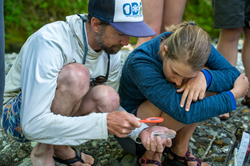
Professor Daryl Teittenen of SNC Tahoe at the river's edge, with a student scientist. Sierra Nevada College Science and Sustainability Department

Professor Daryl Teittenen (left) of SNC Tahoe at the river’s edge, with a student scientist.

By working and learning in the ecosystems that require attention, the RIVER-FSN provides students with education and experience to understand and address the challenges impacting river systems.

SNC Tahoe joins educational consortium to build RIVER Field Studies Network

Freshwater systems are under threat. Non-Native plants and animals, nutrient pollution, growing population pressures, and newly-identified concerns like microplastics – this is just the shortlist of diverse and often synergistic pressures our freshwater resources face. To help student-scientists of the future address these issues, Dr. Andy Rost, of Sierra Nevada College, joins forces with Dr. James Vonesh of Virginia Commonwealth University and Dr. Danielle Perry of Northern Arizona University through a grant from the National Science Foundation. Drs. Rost, Vonesh and Perry will work with seven other researchers from around the country. Their mission? As principal investigators, build a framework for the River-based Immersive Education and Research Field Studies Network (RIVER-FSN). The RIVER-FSN takes some of its cues from the Organization for Tropical Studies, focusing on Undergraduate Biology Education. The network’s participants intend to guide new development of river-based field studies curriculum and programs.

Building a network for collaborative and comparative research

By working and learning in the ecosystems that require attention, the RIVER-FSN provides students with education and experience to understand and address the challenges impacting river systems. Using these freshwater systems as field sites, Vonesh, Perry, and Rost are building a network of researchers, trainers, and collaborators. Once complete, the RIVER-FSN will employ active site experiences and build broader connections throughout the field of undergraduate biology curriculum.

A vital system for field experience

The network and incubator, its capacities, and challenges are vital for many reasons. Rivers are natural classrooms for STEM learning, with processes that integrate biology, hydrology, geology, human, and natural systems. Rivers also provide essential services to society at large, while being among the most imperiled ecosystems. Because of these and many other features, they offer unique solutions for many barriers to student field studies.

Engaged students and the communities they learn in

“Engaging students in expeditionary research holds exceptional promise for biology education due to the breadth and importance of river research, the immersive nature of river expeditions, and the use of high-impact active learning pedagogy,” states their project overview. As an incubator project, RIVER-FSN seeks to bring together a community already invested independently in river-based field studies and expands opportunities for collaborative research and information sharing.

Funding to build a network of active learning

The RIVER-FSN builds capacity for active-learning environments that also includes comparative river biology. Beginning with a series of workshops, the project will identify topics, create expectations, and establish communications channels. The first of these workshops takes place on the American River, hosted by Dr. Rost and professor Daryl Teittenen of SNC Tahoe. Jointly funded by the Directorate for Biological Sciences, Division of Biological Infrastructure, and the Directorate for Education and Human ResourcesDivision of Undergraduate Education, the RIVER-FSN is part of the effort to address the challenges posed in Vision and Change in Undergraduate Biology Education: A Call to Action (http://visionandchange.org/finalreport/).

Share article on social media or email:
For the First Time, UMD Professor Observes Crystallized Iron Product, Hemozoin, Being Made in Mammals, with Widespread Implications for Treatment of Blood Disorders


Professor Iqbal Hamza

Professor Iqbal Hamza, Credit: Edwin Remsberg

Blocking the heme transporter gene transiently should be able to overcome acute heme toxicity in diseases such as sickle cell and malaria where the cells become significantly injured, causing pain, says Hamza.

Most people have some idea of how important iron is to their health, but Iqbal Hamza, professor in Animal and Avian Sciences, has made his seventeen-year career at the University of Maryland all about the study of iron and heme trafficking and regulation in the body. With his latest publication in eLife, Hamza has discovered a never-before-seen protection mechanism in mammals against the toxicity of free heme in the body – the production of a crystallized form of heme known as hemozoin. The production of hemozoin was previously thought only to be possible by blood-feeding organisms like those that cause malaria, but observing this protective phenomenon in mammals opens up entirely new lines of research into how heme tolerance occurs in humans and how this can be used to treat not only malaria and other parasitic infections, but also hemolytic diseases like sickle cell disease.

“Every second, our body recycles over 1000 trillion heme-iron obtained from breaking down hemoglobin from dying red blood cells,” says Hamza. “The gene that regulates heme transport is highly expressed in the spleen, liver, and bone marrow. Specialized macrophages help nurse and make new red blood cells, but they also help clear out the old ones. In fact, 5 million red blood cells are dying every second from age. These macrophages then help to recycle the heme-iron and take it back to the bone marrow. This is the heme-iron recycling process, and it is happening at an astonishing rate every second. But this study shows that mutating the main heme transporter in mice blocks heme recycling, and instead of this being lethal, it forms hemozoin. This has never been seen in mammals before.”

Hemozoin is a crystallized form of heme that is much less toxic and damaging to the system. Hamza previously identified the gene (known as HRG1) that regulates heme transport in his lab, first using a model organism called Caenorhabditis elegans, a bloodless worm that does not create its own heme like mammals do, making it easier to study and track heme transport in their bodies without getting confused as to what heme is coming from where. He then identified a similar gene in humans and wanted to test its function by knocking it out in a mammal to confirm its role in heme transport and recycling. Mouse genetics were performed in collaboration with David Bodine, senior investigator in the Genetics and Molecular Biology Branch at the National Human Genome Research Institute (NHGRI), an institute of the National Institutes of Health (NIH). This study not only confirmed the role of the gene in heme transport, but led to the discovery that mammals will find a way to survive under these conditions through the production of hemozoin.

“Heme-iron recycling throughout the body is like a constant flow of water in a looped system,” says Hamza. “If you mess up one step, you can clog the system and cause build up. Without hemozoin formation, that heme build up would and should be lethal because free heme is so toxic. And even intuitively, we would have expected these mice to be significantly impaired. But the mice don’t show any kind of obvious impairments and look healthy from the outside. Up till now, any mammal who manages to accumulate heme ends up dying because heme is toxic. So how can these animals become heme tolerant?” Heme and iron measurements were done in collaboration with John Phillips, professor of pathology from the Division of Hematology, University of Utah School of Medicine.

Furthermore, if this phenomenon is being observed in mice, Hamza is “willing to bet” it is also occurring in humans. “Heme-tolerant patients could be mistaken as malaria patients, because when hemozoin is found in a human subject, it is assumed that the hemozoin is being made by the malaria parasite. We didn’t know that mammals could make their own hemozoin until now.”

Hamza continues, “I bet that there must be humans with hemozoin in their liver, spleen, and marrow, and it is only a matter of time before we find them. Our data predicts that these people will be protected from heme toxicity. We have to actively go and look for these patients so we can study the mutation of this gene in humans, because they will have less severe ailments than what we would typically expect.”

But finding these patients is important for the future study of this mutation and for potential therapeutic functions, as this mechanism could be used to provide relief for patients that suffer from diseases like sickle cell disease or malaria.

“Blocking the heme transporter gene transiently should be able to overcome acute heme toxicity in diseases such as sickle cell and malaria where the cells become significantly injured, causing pain. This would create some hemozoin in the body, but the animals should survive giving them extra time to reprogram themselves to deal with heme toxicity. This could have an important therapeutic advantage during hemolysis.”

Beyond this, there are a slew of other research questions that are opened up and can now be explored thanks to this discovery in mammals. The mechanisms underlying how hemozoin is made and how heme tolerance occurs are unknown, and being able to answer these questions in a human cell are important to the future of this work. Hamza has already had a lot of interest in this work, with collaborators coming out of fields from immunology to parasitology to neuroscience to microbiology. There are even urologists who think this pathway may be important in urinary tract infections and want to collaborate with Hamza.

“The discovery is very exciting, and I’ve gotten several calls already about all the different directions we can take this,” says Hamza. “It’s great to see the support in the scientific community, and we are already planning to search for this in humans to continue the path towards treatment of parasitic diseases, hemolytic diseases, like sickle cell, and many others.”

The paper entitled, “Hemozoin produced by mammals confer heme tolerance,” is available at eLife, DOI: 10.7554/eLife.49503.

Additional collaborators:

X-ray fluorescence microscopy was performed in collaboration with Martina Ralle, assistant professor in the Department of Molecular and Medical Genetics, Oregon Health and Science University. Macrophage analyses were performed in collaboration with Dr. Malay Haldar, assistant professor of pathology in the Department of Pathology and Laboratory Medicine, University of Pennsylvania. X-ray powder diffraction analyses were performed in collaboration with Dr. Eiji Nishibori from University of Tsukuba, Japan and Hiroshi Sugimoto, structural biologist from RIKEN SPring-8 Center, Japan.

Share article on social media or email:

Spend Matters Rates APEX Analytix as the Top Supplier Relationship Management and Risk Solution


APEX Analytix

We are proud to provide solutions that support 100% of suppliers, regulations and policies in a central supplier dashboard.

APEX Analytix has been recognized as the customer and analyst leader in Supplier Relationship Management and Risk solutions by Spend Matters in its Q3 2019 SolutionMap for procurement technology. This is the second consecutive quarter that APEX Analytix received the top ratings from both customers and analysts. Well-respected as a snapshot of the best solutions in the market, the SolutionMap provides customer and analyst scoring across 5 buyer personas, 56 procurement software companies and 12 solution categories.

“Our clients demand what the ‘end-to-end’ Source-to-Pay suites have never been able to deliver,” describes Akhilesh Agarwal, SVP, Global Business Partner Technology Solutions at APEX Analytix. “We are proud to provide solutions that support 100% of suppliers, regulations and policies in a central supplier dashboard.”

Designed to engage every supplier, the self-service APEX Portal is the fastest growing supplier portal in Source-to-Pay history with 3 million suppliers in 3 years. Meaningful financial impact is derived by addressing the complete supplier lifecycle, validating supplier data with APEX Analytix’s SmartVM® software, and seamlessly integrating with other Source-to-Pay solutions.

Unlike any data offering in the market, SmartVM® validates and enriches supplier data through integration with more than 650 government, regulatory and trusted third-party data sources and over 32 million golden records for suppliers to the Global 2000.

“Beyond feature and function, we look at the platform elements to see what’s real and what’s not,” said Pierre Mitchell, Chief Research Officer at Spend Matters. “Some users look for configurability, others don’t. We reflect buyer needs against, for instance, the application of AI, user experience and low-code platforms. With SolutionMap, procurement has this information at their fingertips.”

“In collaboration with the world’s most advanced Source-to-Pay organizations, we’ve created the first supplier management solution for every supplier,” says Steve Yurko, CEO of APEX Analytix. “We are excited to have this independent validation that we’re delivering solutions that meet customer needs.”

About Spend Matters

Spend Matters is the leading solution intelligence source for procurement and supply chain professionals. Combining deep technology analysis and tailored advisory services with daily news coverage and subscription research, Spend Matters is trusted by CPOs, consultants, investors and solution providers alike as their procurement technology intelligence partner. Spend Matters is owned and managed by Azul Partners Inc.

About APEX Analytix

APEX Analytix revolutionized recovery audit with advanced analytics and the introduction of FirstStrike® overpayment prevention software in 1998. Today, APEX Analytix leads the world in supplier management innovation with APEX Portal and SmartVM, the most popular supplier onboarding and compliant master data management solutions available. With over 250 clients in the Fortune 500 and Global 2000, APEX Analytix is dedicated to providing companies and their suppliers the ultimate supplier management experience. Learn more at http://www.apexanalytix.com.

Share article on social media or email:
Diablo magazine writes about UCSF Benioff Children’s Hospital Oakland and its innovative use of virtual reality technology in the October 2019 issue.


"Virtual Healing" illustration by Stefania Infante

“Virtual Healing” article featured in Diablo magazine October 2019

“It’s very empowering, because you can see things that were otherwise hidden,” Dr. Auguste says, “It makes me think back to all of the difficult surgeries I’ve ever done. If only I’d had this technology before, I wonder how much easier those experiences might have been.”

Dr. Kurtis Auguste is a pediatric neurosurgeon at UCSF Benioff Children’s Hospital Oakland, and he uses virtual reality (VR) technology to prepare and plan for his surgeries. When compared to the two-dimensional, thin, vertical segments of an MRI, the 3D replica of the entire area in question is a game changer. Now, he is sharing that technology with patients and their families prior to surgery to educate and reassure them. Read the entire story here.

Using a virtual reality system called Surgical Theater, Dr. Auguste can put on a headset and “fly” into a 3D model of a young patient’s brain. The patient and his or her parents are invited to a pre-operative session where they all wear goggles and are able to watch and individually interact with the visual experience. Dr. Auguste, represented by an avatar wearing a white doctor’s coat, serves as tour guide. Using hand controls to glide around, they can see blood vessels, vision fibers, and even tumors or aneurysms. Dr. Auguste plots his point of entry for surgery and how best to remove threatening tissue abnormalities. Patients and their families benefit from a visual introduction to the cause of the patient’s medical condition and the knowledge of exactly how Dr. Auguste intends to operate on it. This exercise brings great calm and confidence to patients, helping them focus on a successful surgery and future recovery.

“It’s very empowering, because you can see things that were otherwise hidden,” Dr. Auguste says, “It makes me think back to all of the difficult surgeries I’ve ever done. If only I’d had this technology before, I wonder how much easier those experiences might have been.”

Dr. Auguste sees many applications for the future use of VR technology in the medical field, including but not limited to educating patients and medical staff, serving as a rehab and recovery monitoring tool, and training international teams of surgeons and medical staff.

Virtual reality also helps young patients manage pain and stress. Patients at UCSF Benioff Children’s can slip on a headset and enter an underwater experience called KindVR Aqua. Simon Robertson, the founder of KindVR in Alameda, says the technology allows patients to interact with dolphins and whales, which introduces a distraction, and helps them reduce the perception of pain and even potential opioid consumption.

ABOUT DIABLO MAGAZINE

Celebrating 40 years, Diablo magazine covers topics ranging from travel, culture, and personalities to entertainment, recreation, and food. Diablo magazine is written specifically for the San Francisco East Bay market—from Central Contra Costa, into the Oakland and Berkeley hills, and throughout the Tri-Valley. With award-winning editorial content and beautiful photography, Diablo magazine is a unique celebration of the San Francisco East Bay. Published since 1979, Diablo magazine has been recognized for its editorial and design with numerous awards, including Maggie Awards for Best Overall Publication and Best Regional and State Magazine in the consumer category. To learn more about Diablo magazine, visit diablomag.com.

ABOUT DIABLO CUSTOM PUBLISHING

Diablo Custom Publishing (DCP), provides print and digital marketing communications and custom publishing services for corporate clients nationwide. DCP is the custom services division of Diablo Publications, which has been an award-winning publisher since 1979. DCP offers marketing expertise and publishing excellence within the fields of healthcare, higher education, visitor travel, and arts & entertainment. To learn more about DCP, visit dcpubs.com.

ABOUT DIABLO PUBLICATIONS

Diablo Publications, an employee-owned company, has been creating award-winning publications, including Diablo magazine, Napa Sonoma magazine, Diablo Weddings, Diablo Arts, Concierge Wine Country, Visit Oakland Inspiration Guide, and the Tri-Valley California Visitors Guide. Diablo Publications’ custom content division, Diablo Custom Publishing (DCP), provides print and digital marketing communications and custom publishing services for corporate clients nationwide. For more information visit diablomag.com to view our retail publications and dcpubs.com to learn more about custom publishing.

Share article on social media or email: